Featured Dental Articles
Clinical Tips and Insights
“In the lead up to our July 2018 symposium, I wish to begin dismantling the misconceptions surrounding Sleep Bruxism and the unsupported views held by some local dentists regarding its management. This is a series of weekly blogs covering various aspects of clinical practice and I hope the insights I have gained over the course of my career, will encourage you to re-evaluate commonly accepted, non-evidence based dogma so prevalent in dentistry.”
I’d like to introduce you to a practical 6-week diagnostic protocol I’ve developed over the years to help me better understand the patient’s bruxism profile or phenotype.
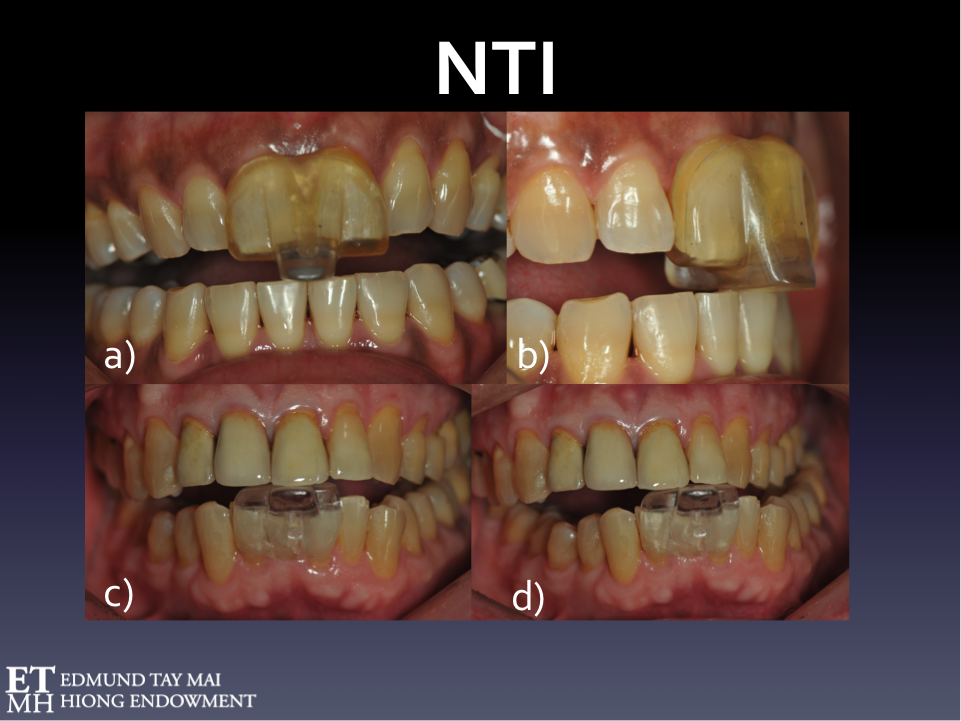
Nociceptive Trigeminal Inhibition Tension Suppression System (NTI-tss)
I have been using the NTI device since 1998. The NTI-tss is an easily customizable, commercially available segmental intraoral device that engages the Trigeminal jaw-opening reflex - a polysynaptic relay involving interneurons, nociceptors and mechanoreceptors in the periodontal ligament. (Fig. 12) Despite providing arguably minimal supportive clinical research evidence[1] the manufacturer managed to gain FDA approval for its use in the treatment of bruxism, TMD, occlusal trauma as well as the prophylactic non-pharmaceutical management of migraine and tension-type headaches.
In today’s blog we continue to explore the different bruxism phenotypes.
Perhaps one of the biggest reasons for the confusion in the literature was the failure to differentiate sleep bruxism from bruxism occurring when awake.
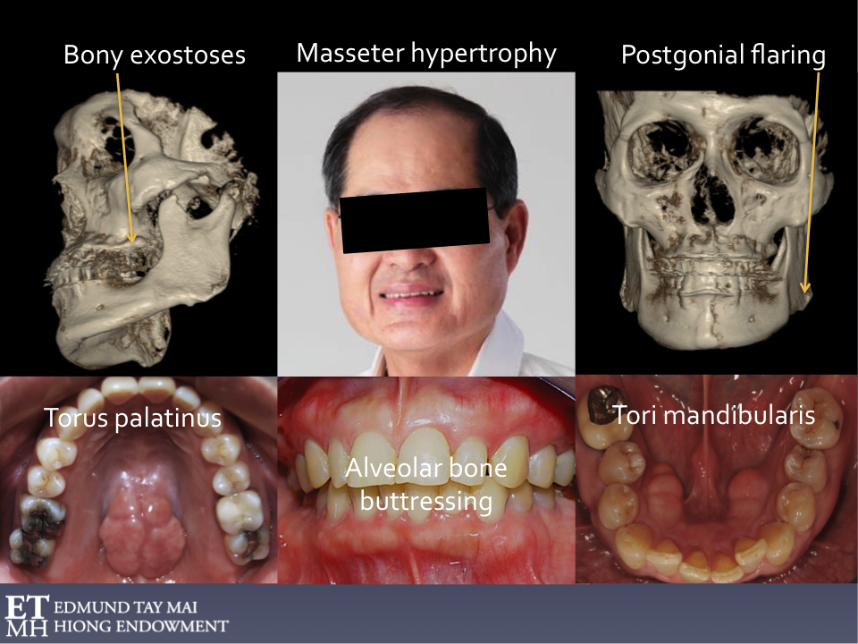
Approximately one third of patients with SB also exhibit concomitant wake-time bruxism. The latter has an estimated prevalence of 12% in children and >20% in adults [1]. Psychosocial factors like stress, anxiety-hypervigilance and personality traits have significant influence on AB whereas there is less evidence to support their role in the pathogenesis of SB. The Polyvagal Theory (Porges, 1995)[2] which describes the development of the mammalian social engagement system and the involvement of the myelinated vagus, provide insights that suggest AB may be an effective adaptive strategy employed by certain individuals under stress to increase their ‘vagal brake’. The stoic socio-cultural environment (rewarding self-control, discouraging the open expression of emotions, keeping your mouth shut and your teeth together!) rather than genetics are thought to be responsible for significant racial differences in the higher prevalence of oral tori/bony exostoses in Asians [3, 4].
In the last blog we highlighted some real world limitations in the use of PSG for diagnosing bruxism in clinical dental practice. However, without the benefit of evidence–based PSG research we will still be in the dark.
Dentists generally become involved only after ‘the rubber hits the road’ i.e. when there’s observable occlusal interface and/or other stomatognathic damage (e.g. TM joint dysfunction, masticatory muscle symptomology). Most are blind to what comes before! (Fig. 3) However, the ability to subcategorize bruxism into different phenotypes according to the brain state in which it occurs is extremely important when tailoring treatment for these patients. Moreover, we need to know if we’re, in fact, dealing with secondary bruxism.
“If it is measured it is a fact, otherwise it is an opinion.” Barney Jankelson
But how it is measured is as important!
Dentistry is fast changing and we all struggle to keep up. What I was taught about bruxism as a student in dental school in the late seventies and what I taught when I was faculty had expiry dates. Recently, I had the privilege to be involved in the setting up of the NUS Edmund Tay Mai Hiong (ETMH) endowed fund which I hope will serve as a useful platform to introduce sleep medicine and airway prosthodontics to the local dental profession.